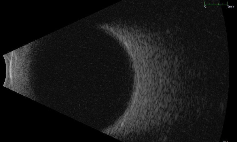
What is an ocular ultrasound?
While the OCT scans use light wavelengths to capture abnormalities present in the macula, ultrasounds (B-scan) uses sound waves to evaluate the overall structural integrity of the posterior segment including the retina.

Why do I need a B-scan?
The B-scan serves as a diagnostic tool that can reliably diagnose hidden pathologies present in the posterior segment of the eye that are otherwise difficult to see during an ocular examination. When view of the retina is obstructed by conditions such as cataracts, small pupil, or internal hemorrhaging, B-scan is helpful in detecting underlying disorders such as retinal tear or detachment, ocular tumor or choroidal lesion.

What to expect when undergoing ocular ultrasonography?
B-scan imaging is a non-invasive process that can take as little as 1 minute. Once seated, the patient will be asked by our technicians to close their eyes as this is performed over the eyelids. The technician will then gently place a probe with sterile gel solution on the eyelid in order to obtain multiple scans of the eye. Once the pictures are taken, they will be uploaded to the patient’s chart where the doctor can review them with patients in the private exam room.
