What is a fundus?
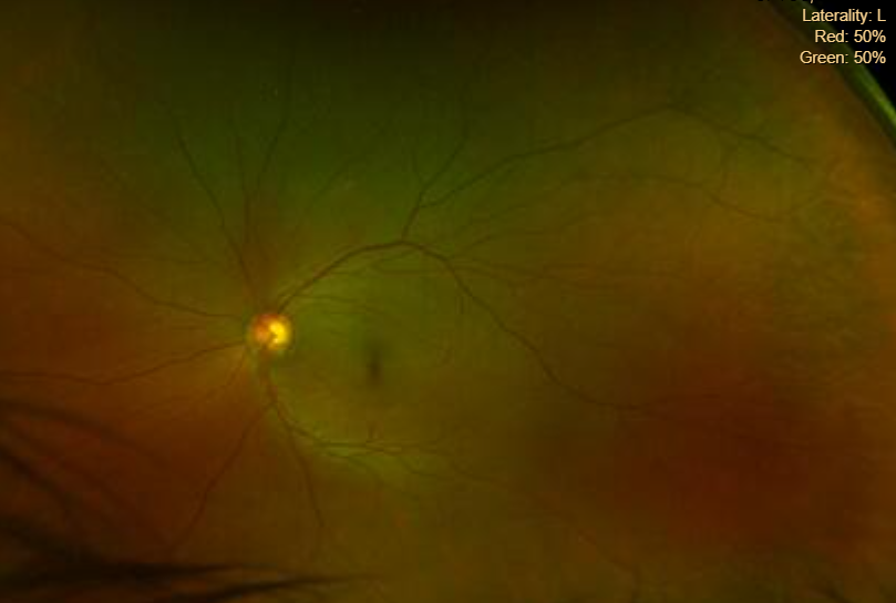
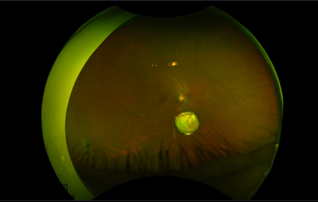
Fundus is a term that is used to describe the inner lining of the back part of the eye (retina). When we perform a digital fundus photography, our trained/experienced photographer is conducting a diagnostic procedure which uses a highly specialized microscope to comprehensively document different parts of the posterior segment (retina, macula, optic nerve) with high-resolution.

Why is a fundus examination done?
The fundus exam helps to capture and document any abnormalities present in the eye. It helps in collecting an accurate medical record of the retina which can be used for comparisons on future examinations to assess any overall change. This is why it is a normal occurrence for returning patients to undergo subsequent fundus imaging to help highlight the progression or management of a particular ocular disease.

What to expect when undergoing fundus photography?
Fundus diagnostic imaging is a fast and non-invasive process that usually takes a minute. Before taking a fundus image with our skilled photographer, patients will be asked to have their eyes dilated. This helps ensure that the view to the retina is optimal. During the test, patients will be sitting comfortably in front of the machine with their chin rested on the chin-rest. Once the pictures are taken, they will be uploaded to the patient’s chart where the doctor can review them with patients in the private exam room.
