What is a detached retina?
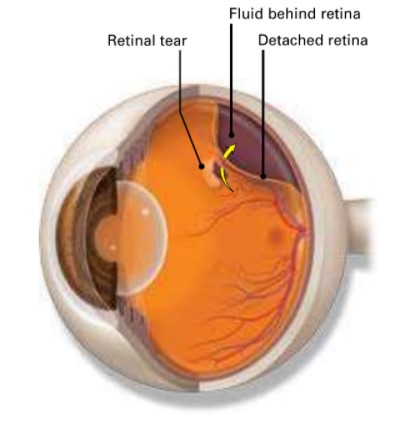
A detached retina is when the retina lifts away from the back of the eye. The retina does not work when it is detached, making vision blurry. A detached retina is a serious problem.
An ophthalmologist needs to check it out right away, or you could lose sight in that eye.
How do you get a detached retina?
As we get older, the vitreous in our eyes starts to shrink and get thinner. As the eye moves, the vitreous moves around on the retina without causing problems. But sometimes the vitreous may stick to the retina and pull hard enough to tear it. When that happens, fluid can pass through the tear and lift (detach) the retina.
® Eye Words to Know
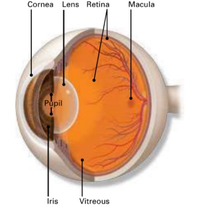
Retina: Layer of nerve cells lining the back wall inside the eye. This layer senses light and sends signals to the brain so you can see.
Vitreous: Jelly-like substance that fills the middle of the eye.
Floaters: Tiny clumps of cells or other material inside the vitreous. These look like small specks, strings or clouds moving in your field of vision.
Who is at risk for a detached retina?
You are more likely to have a detached retina if you:
- need glasses to see far away (are nearsighted)
- have had cataract, glaucoma, or other eye surgery
- take glaucoma medications that make the pupil small (like pilocarpine)
- had a serious eye injury
- had a retinal tear or detachment in your other eye
- have family members who had retinal detachment
- have weak areas in your retina (seen by an eye doctor during an exam)
Early signs of a detached retina
A detached retina has to be examined by an ophthalmologist right away. Otherwise, you could lose vision in that eye. Call an ophthalmologist immediately if you have any of these symptoms:
- Seeing flashing lights all of a sudden. Some people say this is like seeing stars after being hit in the eye.
- Noticing many new floaters at once. These can look like specks, lines or cobwebs in your field of vision.
- A shadow appearing in your peripheral (side) vision.
- A gray curtain covering part of your field of vision.
How is a detached retina diagnosed?
Your ophthalmologist will put drops in your eye to dilate (widen) the pupil. Then they will look through a special lens to check your retina for any changes.
How is a detached retina treated?
Surgery is done to repair a detached retina. Here are some types of detached retina surgery:
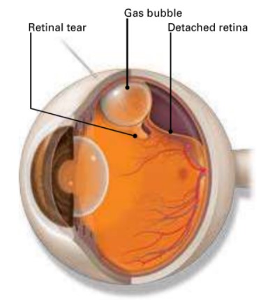
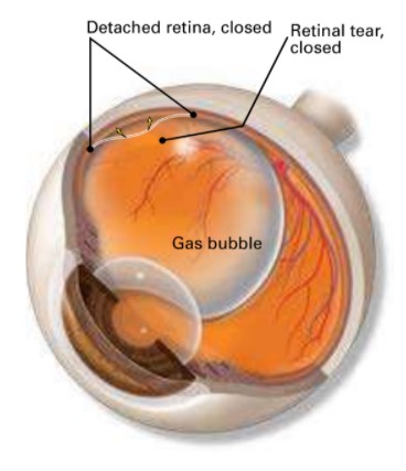
Pneumatic retinopexy. Your ophthalmologist puts a gas bubble inside your eye. This pushes the retina into place so it can heal properly. Afterwards, you will need to keep your head in a very specific position as your doctor recommends for a few days. This keeps the bubble in the right place. As your eye heals, your body makes fluid that fills the eye. Over time, this fluid replaces the gas bubble.
Vitrectomy. Your ophthalmologist removes the vitreous pulling on the retina. The vitreous will be replaced with an air, gas, or oil bubble. The bubble pushes the retina into place so it can heal properly. If an oil bubble is used, your ophthalmologist will remove it a few months later. With an air or gas bubble, you cannot fly in an airplane, travel to high altitudes or scuba dive. This is because altitude change causes the gas to expand, increasing eye pressure.
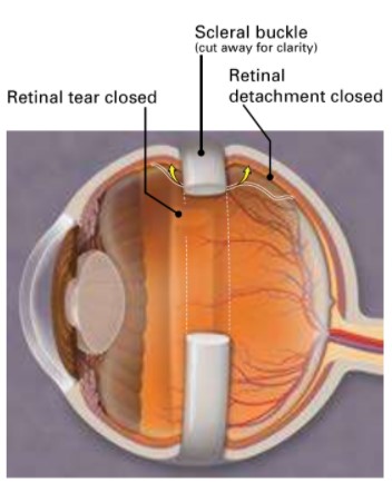
Scieral buckle. A band of rubber or soft plastic is sewn to the outside of your eyeball. It gently presses the eye inward. This helps the detached retina heal against the eye wall. You will not see the scleral buckle on the eye. It is usually left on the eye permanently.
What are the risks of surgery for detached retina?
All surgery has risks of problems. But if you do not treat a detached retina, you could quickly and permanently lose your sight. Here are some of the risks of surgery for detached retina:
- Eye infection
- Bleeding in your eye
- Glaucoma, when pressure increases inside the eye
- Cataract, when the lens in your eye becomes cloudy
- Need for a second surgery
- Chance that the retina does not reattach properly
- Chance that the retina detaches again
Your ophthalmologist will discuss these and other risks and how surgery can help you.
Things to expect after surgery:
- You might have some discomfort for a few days to weeks after surgery. You will be given pain medicine to help you feel better.
- You need to rest and be less active after surgery for a few weeks. Your ophthalmologist will tell you when you can exercise, drive or do other things again.
- You will need to wear an eye patch after surgery. Be sure to wear it as long as your doctor tells you to.
- If a bubble was put in your eye, you will need to keep your head in one position for a certain length of time, such as 1-2 weeks. Your doctor will tell you what that specific head position is. It is very important to follow the directions so your eye heals.
- You might see floaters and flashing lights for a few weeks after surgery. You may also notice the bubble in your eye.
- Your sight should begin to improve about four to six weeks after surgery. It could take months after surgery for your vision to stop changing. Also, your retina may still be healing for a year or more after surgery. How much your vision improves depends on the damage the detachment caused to the cells of the retina.
Summary
A detached retina is when your retina lifts away from the back of the eye. You may see flashing lights or many new floaters. Or you may see a shadow in your side vision. Also, a gray curtain might cover part of your field of vision. If you have any of these signs, call an ophthalmologist right away.
Surgery may be done to help your retina heal in place against the eye wall.
A type of bubble may be placed in your eye to help the retina heal in place. You must follow your ophthalmologist’s instructions after surgery to help make sure your eye heals properly.
If you have any questions about your vision, speak with your ophthalmologist. He or she is committed to protecting your sight.
Watch a detached retina video from the American Academy of Ophthalmology’s EyeSmart program at aao.org/detached-retina-link.
