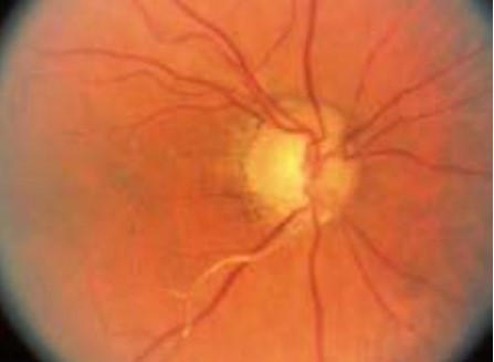
What is a retinal artery occlusion?
Most people know that high blood pressure and other heart diseases pose risks to your overall health. But many do not know that that high blood pressure can affect vision by damaging the arteries in the eye.
A retinal artery occlusion (RAO) is a blockage in one or more of the arteries of your retina. The blockage is caused by a clot or occlusion in an artery, or a build-up of cholesterol in an artery.
There are two types of RAOs:
- Branch retinal artery occlusion (BRAO) blocks the small arteries in your retina.
- Central retinal artery occlusion (CRAO) is a blockage in the central artery in your retina. This is a form of a stroke in the eye and must be treated immediately.

What are symptoms of an RAO?
The most common symptom of an RAO is sudden, painless vision loss. It can affect all of one eye, in the case of CRAO, or it can affect part of one eye, in the case of BRAO. Other symptoms include:
- loss of peripheral vision
- distorted vision
- blind spots
If you have any of these symptoms, get medical help right away to help prevent vision loss.
® Eye Words to Know
Retina: Layer of nerve cells lining the back wall inside the eye. This layer senses light and sends signals to the brain so you can see.
Occlusion: The narrowing or closure of a blood vessel that carries blood to the retina.
High blood pressure: This is measured by the amount of blood flow in your body and your resistance to blood flow. If your heart pumps a lot of blood and you have very narrow arteries, you have high blood pressure.
Who is at risk for an RAO?
Men are more likely to have an RAO than women. The disease is most commonly found in people in their 60s. Having certain diseases increases your risk of RAO. These include:
- cigarette smoking
- cardiovascular (heart) disease
- diabetes
- high cholesterol
- high blood pressure
- narrowing of the carotid or neck artery

How is RAO diagnosed?
If you experience sudden vision loss, you should contact your ophthalmologist immediately. He or she will conduct a thorough examination to determine if you have had an RAO. Your ophthalmologist will dilate your eyes with dilating eye drops. This will allow him or her to examine the retina for signs of damage.
Other tests your ophthalmologist may do are:
- fluorescein angiography. This imaging test uses a special camera to take photographs of the retina. A small amount of yellow dye
(fluorescein) is injected into a vein in your arm. The photographs of fluorescein dye traveling throughout the retinal arteries show how many arteries are closed.
intraocular pressure (pressure inside the eye) - reflexes of your pupil
- other photos of the retina
- a slit-lamp examination
- testing of side vision (visual field examination)
- visual acuity (sharpness), to see how well you can read an eye chart
Since RAOs represent urgent ophthalmic conditions, they require prompt evaluation. Your ophthalmologist will also likely communicate with your medical provider. The evaluation may include an ultrasound of your carotid arteries (the main blood vessels in your neck that send blood to your eyes and brain). An ultrasound is a medical test that uses soundwaves to create images of the organs and tissues inside your body. You might also be told to have an echocardiogram (ultrasound of the heart). Both of these tests help look for a possible source of the blockage in the retinal artery. In most cases of CRAO, a prompt referral to a stroke center for a medical evaluation is recommended because the risk of stroke is particularly high during the first 1 to 4 weeks.
How is RAO treated?
Several treatments may be tried but none have ever been proven to help consistently. These treatments must be given within a few hours after symptoms begin to be helpful. Treatments include:
- breathing in (inhaling) a carbon dioxideoxygen mixture. This treatment causes the arteries of the retina to widen (dilate).
- removing some liquid from the eye to allow the clot to move away from the retina
- a clot-busting drug
Some patients regain vision after an RAO, although vision is often not as good as it was before. In some cases, vision loss can be permanent.
Summary
A retinal artery occlusion (RAO) happens when there is a blockage of blood flow to the retina in the back of the eye. Symptoms include sudden vision loss, distorted vision or blind spots in your vision. Certain health conditions like diabetes and high blood pressure increase your risk for having an RAO.
If you have symptoms of an RAO, see an ophthalmologist as soon as possible. He or she can look for signs of damage and treat you if necessary. In some cases, patients can regain vision after an RAO. In others, however, vision loss can be permanent. This is why it is so important to see an ophthalmologist or doctor at the first sign of any symptoms.
If you have any questions about your vision, speak with your ophthalmologist. He or she is committed to protecting your sight.
Get more information about retinal artery occlusion from EyeSmart-provided by the American Academy of Ophthalmologyat aao.org/retinal-artery-occlusion-link.
