What is cystoid macular edema?
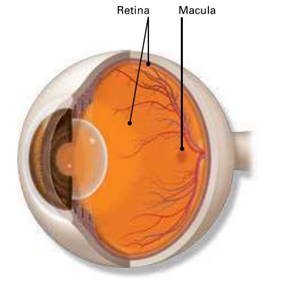
Cystoid macular edema (CME) is an eye disease that affects part of your retina. It is when the macula becomes swollen and tiny blisters of fluid form that look like small cysts.
Layer of cells lining the back wall inside the eye. This layer senses light and sends signals to the brain so you can see.
What are symptoms of CME?
The most common symptom of CME is distorted central vision. This is when things in front of you look wavy or blurry. CME does not affect your peripheral (side) vision. You may find that objects look dark or dim. Sometimes, things can look pink (even though they are not really pink). You also may be sensitive to light.
You can have CME without any of these symptoms.

Retina: Layer of cells lining the back wall inside the eye. This layer senses light and sends signals to the brain so you see.
Macula: Small but important area in the center of the retina. You need the macula to clearly see details of objects in front of you.
Cysts: Tiny, fluid-filled sacs in the macula.
What causes CME?
You are more likely to have CME if you have had:
- blocked blood vessels in the retina (called retinal vein occlusion)
- uveitis (when the layer of the eyeball under the white of the eye is swollen)
- an eye injury
- CME in the other eye
- diabetes, or if you take certain medicines for glaucoma or diabetes, or take niacin (vitamin B3)
- cataract surgery or other eye surgery.
Your version may start to improve within a few months after having your CME treated.
How is CME dianosed?
Your ophthalmologist will put drops in your eye to dilate (enlarge) the pupil. Then he or she will look at the back of your eye through a special lens. This allows your doctor to see if there are changes to the retina and macula.
Your doctor may do to see what is happening with your retina. Yellow dye (called fluorescein) is injected into a vein, usually in your arm. The dye travels through your blood vessels. A special camera takes photos of the retina as the dye travels throughout its blood vessels. This shows any changes to the retina or macula.
Optical coherence tomography (OCT) is another way your ophthalmologist can look closely at the retina. A special type of machine scans the retina and provides very detailed images of the retina and macula.
How is CME treated?
There are many ways to treat CME. They include:
- eye drops to reduce swelling in the macula. These drops could be steroid or non-steroid eye drops
- injections (shots) of medicine in the eye
- other types of drugs to reduce swelling
- laser surgery to repair leaking blood vessels in your retina
- surgery called vitrectomy to remove scar tissue on your macula
Your ophthalmologist will talk about which treatment he or she recommends for you.
Your vision may start to improve within a few months after having your CME treated.
Summary
Cystoid macular edema (CME) is where tiny, fluid-filled sacs called develop in your macula. It affects your central vision. It may be treated with eye drop medicines, medication injections, laser surgery, or other surgery.
If you have any questions about your eyes or your vision, speak with your ophthalmologist. He or she is committed to protecting your sight.
